Test Prep for AP® Courses


- Natural killer cells recognize MHC I on a healthy cell and do not kill it, while the infected cells that do not present MHC I are killed.
- Natural killer cells recognize MHC I on an infected cell and kill it, while the healthy cells that do not present MHC I are not killed.
- Natural killer cells recognize MHC II on a healthy cell and do not kill it, while the infected cells that do not present MHC II are killed.
- Natural killer cells recognize MHC II on an infected cell and kill it, while the healthy cells that do not present MHC II are not killed.
The incomplete diagram represents a series of events during an innate immune response. The labels A, B, and C need to be replaced with the names of the cells involved.

Which set of cells correctly completes this diagram?
- A = infected host cell, B = pathogen, C = healthy host cell
- A = healthy host cell, B = pathogen, C = dendritic cell
- A = dendritic cell, B = infected host cell, C = pathogen
- A = pathogen, B = dendritic cell, C = healthy host cell
- CD4+T cells are a required intermediate in a series of cell-to-cell signaling events that must be completed before B cells can mature.
- CD4+T cells have CD4 molecules covalently bound to their cell surfaces and do not induce apoptosis in other cells during an immune response.
- CD4+T cell counts are about 1,000 per microliter in a healthy person, but drop below 400 per microliter in a person who cannot mount an immune response.
- CD4+T cell precursors are formed in the bone marrow and then migrate to the thymus, where they develop their T cell receptors.


The graph shows changes in a person’s blood after he or she receives a vaccination.

Explain how cell communication is involved in bringing about the changes depicted in the graph.
- The vaccine introduces antigens specific to a pathogen into the person’s blood. These antigens are moved to the cell surface of antigen-presenting cells present in the blood. Receptors on helper T cells bind to the antigens present on the antigen-presenting cell. This direct cell-to-cell contact initiates a series of events that leads to production of antibodies by B lymphocytes.
- The vaccine introduces antigens specific to a pathogen into the person’s blood. These antigens bind to the receptors on the surface of T cells. This direct cell-to-cell contact initiates a series of events that leads to production of antibodies by B lymphocytes.
- The vaccine introduces antigens specific to a pathogen into the person’s blood. These antigens are moved to the cell surface of antigen-presenting cells present in the blood. This direct cell-to-cell contact initiates a series of events that leads to production of antibodies by B lymphocytes.
- The vaccine introduces antigens specific to a pathogen into the person’s blood. These antigens are moved to the cell surface of antigen-presenting cells present in the blood. Receptors on helper T cells bind to the antigens present on the antigen-presenting cell. This direct cell-to-cell contact initiates a series of events that activates the complement system.
- T cells secrete cytokines, which help the B cell to multiply and mature into an antibody-producing plasma cell.
- Natural killer cells secrete cytokines, which help the B cell to multiply and mature into an antibody-producing plasma cell.
- T cells secrete interferons, which help the B cell to multiply and mature into an antibody-producing plasma cell.
- Natural killer cells secrete interferons, which help the B cell to multiply and mature into an antibody-producing plasma cell.
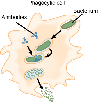
The diagram illustrates a process taking place during an immune response.

What process is represented by this diagram?
- opsonization
- apoptosis
- neutralization
- complement activation
Scientists performed an experiment using a cell from a mouse embryo and a B cell from an adult mouse. The mouse embryo cell does not make antibodies, yet its DNA contains nucleotide sequences encoding antibody polypeptides. The adult mouse B cell makes and secretes a single type of antibody. In the experiment, radiolabeled DNA probes were synthesized to be complementary to the DNA encoding the light chains of the antibody produced by the adult mouse B cells. Then, DNA from the mouse embryo cell and from the adult B cell were isolated and tested to see if either hybridized with the synthesized radiolabeled DNA probes. The results are shown in the diagram.
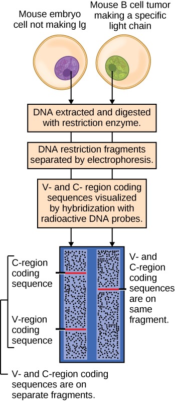
Which claim is best supported by the data?
- The mouse genome contains an enormous number of antibody genes, which accounts for the huge diversity of antibody molecules that can be made.
- Rearrangement of gene segments encoding antibody polypeptides occurs at the level of DNA to produce an enormous diversity of antibody molecules.
- The tremendous diversity of antibody molecules that can be made results from post-translational modifications of antibody polypeptide chains.
- Each antibody is encoded by its own unique gene in the DNA, which explains how antibodies can have different antigen binding properties.
How does an antibody molecule bind specifically to one antigen but not to others?
- due to the presence of a specific antigen binding site
- due to the consistency of the constant region
- due to diversity of the constant region
- due to the complete antibody structure
- There are so many different antibody molecules that can be made, each of which can specifically target a particular pathogen to destroy it. This specificity makes the immune system more effective. The immune system is also efficient because each antibody need to have its own gene.
- There are so many different antibody molecules that can be made, each of which can non-specifically target a particular pathogen to destroy it. This non-specificity makes the immune system more effective. The immune system is also efficient because each antibody does not need to have its own gene.
- There are so many different antibody molecules that can be made, each of which can specifically target a particular pathogen to destroy it. This specificity makes the immune system more efficient. The immune system is also effective because each antibody does not need to have its own gene.
- There are so many different antibody molecules that can be made, each of which can specifically target a particular pathogen to destroy it. This specificity makes the immune system more effective. The immune system is also efficient because each antibody does not need to have its own gene.
An allergy is caused by the immune system reacting to a foreign protein to produce IgE molecules that recognize the protein. These IgE molecules become associated with mast cells that respond to future exposures to the protein by releasing histamines into the body. The diagram shows this release and also how a drug called an antihistamine can help an allergy sufferer reduce his or her allergy symptoms.

Which of the following statements explains how an antihistamine helps restore homeostasis during an allergic reaction?
- Antihistamines prevent mast cells from becoming associated with IgE molecules that recognize the foreign protein allergen.
- Antihistamines prevent mast cells from releasing histamines and causing the unpleasant allergy symptoms.
- Antihistamines prevent histamines that have been released by mast cells from stimulating the itching and swelling of body tissues.
- Antihistamines prevent mast cells from producing histamines, which halts their effect on the body.
The diagram shows the normal feedback loop that controls the production of thyroid hormones in the human body.

Graves’ disease is an autoimmune disease in which the body produces autoantibodies to the TSH receptor. When these autoantibodies bind to the receptor, it mimics the action of the TSH hormone. How would the feedback loop and the regulated production of thyroid hormones shown in the diagram be affected in a person with Graves’ disease?
- The feedback loop would be disrupted. Autoantibodies would bind to the TSH receptors, allowing them to continue to produce thyroid hormones. As a result, there would be an overproduction of thyroid hormones because the negative feedback system would be unable to function.
- The feedback loop would be not be disrupted. Autoantibodies would bind to the TSH receptors, allowing them to continue to produce thyroid hormones. As a result, there would be an overproduction of thyroid hormones because the negative feedback system would be unable to function.
- The feedback loop would be disrupted. Autoantibodies would not bind to the TSH receptors, allowing them to continue to produce thyroid hormones. As a result, there would be an overproduction of thyroid hormones because the negative feedback system would be unable to function.
- The feedback loop would be disrupted. Autoantibodies would bind to the TSH receptors, allowing them to continue to produce thyroid hormones. As a result, there would be an overproduction of thyroid hormones because the negative feedback system was functional.
Myasthenia gravis is an autoimmune disease that initially presents with muscle weakness and can progress to complete impairment of muscle movement. The diagram compares a healthy individual with an individual suffering from this disease.

Which statement best explains what happens to bring about this disease?
- The body produces antibodies against nerve cells, which prevent the nerve cells from releasing acetylcholine during signal transmissions to muscle.
- The body produces antibodies against acetylcholine, which prevent acetylcholine from transmitting signals from nerves to muscle.
- The body produces antibodies against receptors in muscle, which prevent acetylcholine from binding and completing nerve signal transmission.
- The body produces antibodies against acetylcholine, which prevent acetylcholine from breaking down after signal transmission is complete.